Research
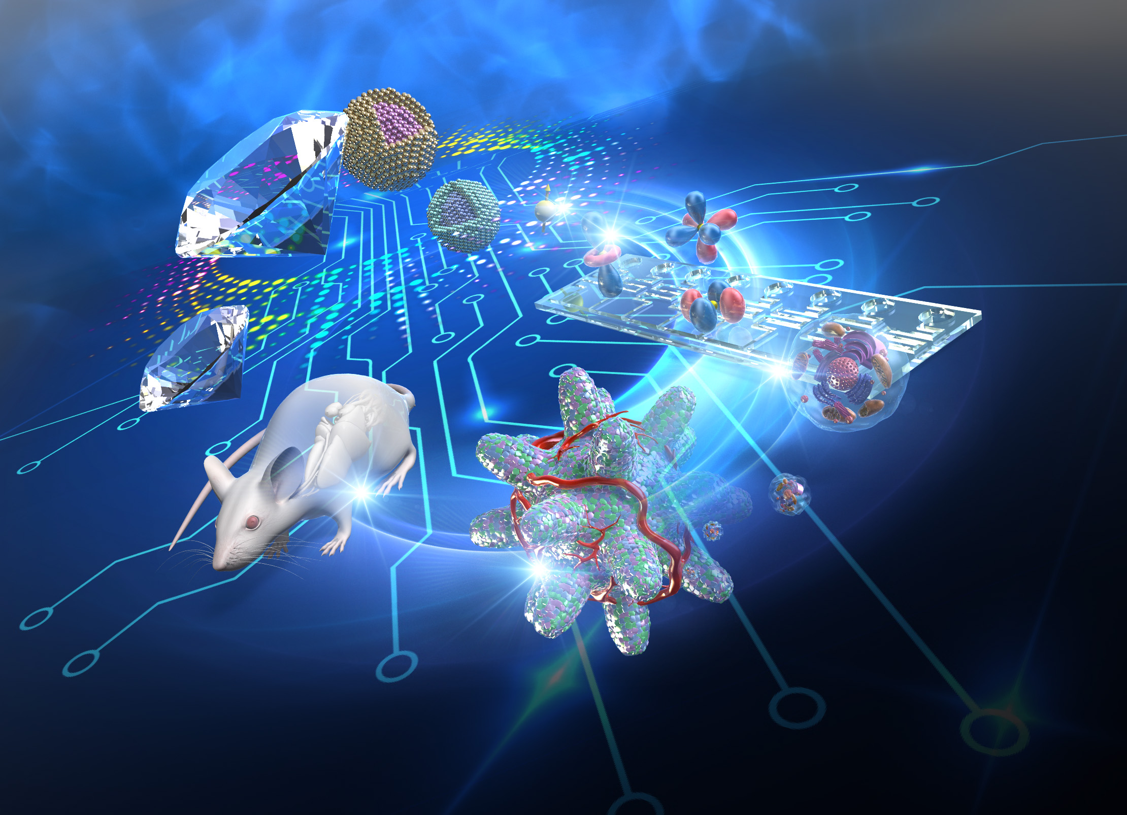
Aiming for Innovation in Regenerative Medicine with Quantum Technology
In the field of regenerative and biomedical engineering, common differentiation induction methods for generating regenerative cells follow embryology and reproduce the concentration, sequence, and reaction time of the addition of regenerative factors. However, information on physicochemical parameters (temperature, pH, radicals, etc.), which largely reflect cellular conditions that change during the actual differentiation process in vivo, is extremely scarce, and this is a major obstacle to establishing differentiation induction methods for cell types that exhibit complex functions.
In regenerative medicine, there is concern that differences in the cellular state of stem cells and regenerated cells may significantly affect the expression of regenerative functions, but the substance of this concern remains elusive. Our team will apply quantum technologies such as nano-quantum sensors to regenerative and biomedical engineering and create technologies to measure physicochemical parameters of cells for quality control of stem cells and regenerative cells before transplantation and for diagnosis in the in vivo environment, tissues, and organs after transplantation. By doing so, we aim to deepen our understanding of the cellular state of stem cells and regenerative cells in vivo and in vitro, and to contribute to the fields of regenerative medicine and embryology.
Development of Nano Quantum sensor and its Application to In Vivo Observation and Diagnosis of Stem Cells and Regenerative Cells
The measurement of "physicochemical parameters" such as temperature, pH, and radicals is important for understanding biological phenomena occurring inside and outside of cells, and there are high expectations for applications such as diagnosis of diseases, development of treatment methods, and cell quality control in regenerative medicine. In particular, if fluctuations in these physicochemical parameters can be detected at high resolution at the single-cell level (measurement of individual cells), it is expected to lead to the creation of methods for ultra-early diagnosis of cancer and evaluation of cell status and function, which are important in regenerative medicine. For example, it is known that inflammation is associated with increases in cell temperature and extracellular pH, and if measurement at single cell resolution can be achieved, it may be possible to detect abnormal cells mixed in a cell population or tissue.
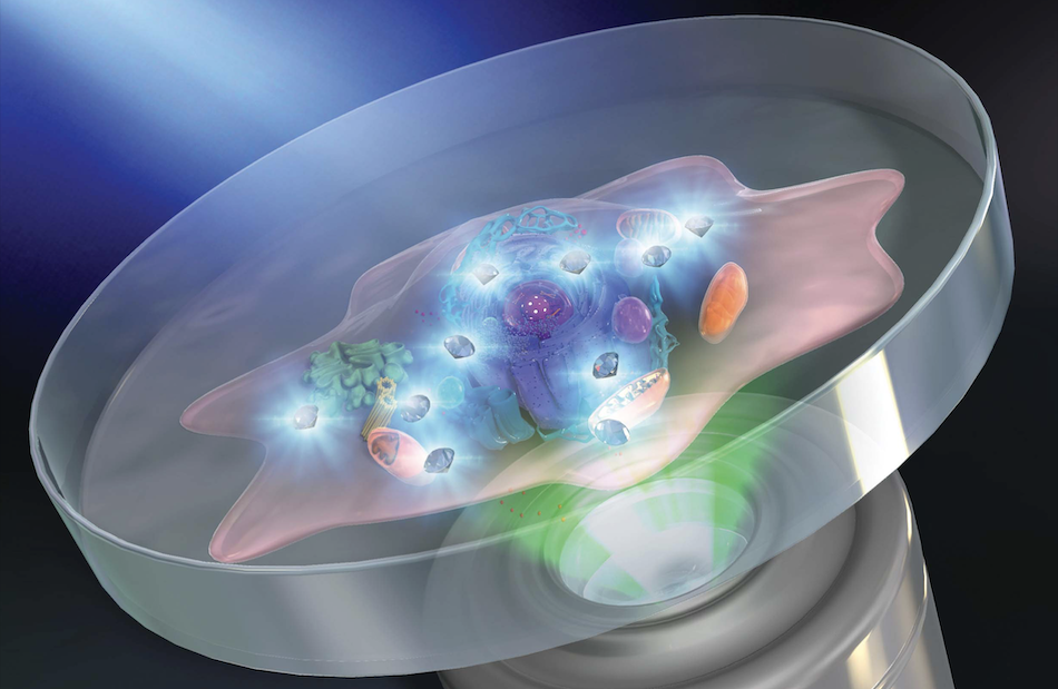
With this in mind, we have developed a single-cell temperature measurement system for stem cells using nano-quantum sensors called "fluorescent nanodiamonds (FNDs)". We found that FNDs incorporated into cells are useful for accurate measurement of intracellular temperature, do not reduce cell viability, and do not inhibit secretion of regenerative factors (e.g., HGF, TGF-β1). In other words, this system enables measurement of cell temperature without affecting stem cell status or functional expression. Through observations using this system, we have also demonstrated that stem cell function is correlated with culture temperature. Furthermore, the application of these techniques to in vivo monitoring is an important issue for the future. Therefore, Dr. Fujiwara's research group (Okayama University) has succeeded in developing a system that can measure temperature inside an individual using the nematode worm, a representative multicellular animal model with the simplest structure.
References
Development of Near-Infrared Nano Quantum Sensor and its Application to In Vivo Deep Imaging Diagnosis and Therapy
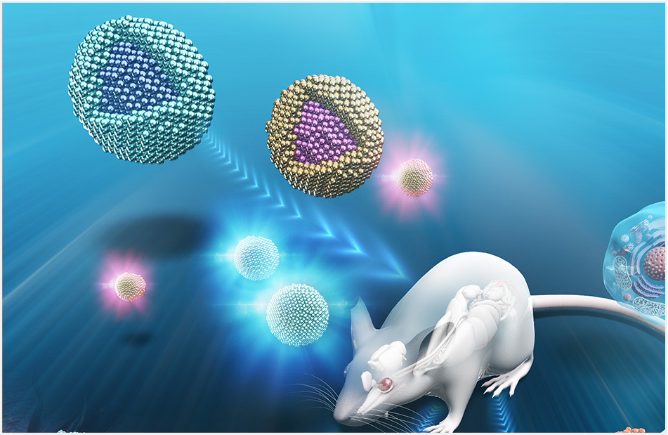
In regenerative medicine, in addition to functional evaluation of stem cells themselves, continuous monitoring of regenerative cells after transplantation into the body is important from the viewpoints of safety and therapeutic efficacy. In parallel with the verification of fluorescent nanodiamond FNDs, we have also been promoting the application of "near-infrared quantum dots (QDs)", which have a proven track record as effective near-infrared fluorescent probes in vivo, to regenerative medicine.
We have already succeeded in developing and commercializing FluclairTM reagents, ultra-low toxicity QDs for stem cell labeling, and we plan to further commercialize these reagents by taking advantage of the diversity of QDs composition to synthesize QDs with various fluorescence wavelength ranges. In particular, the use of low-toxicity QDs that show strong fluorescence in the near-infrared region (around 700-900 nm), which is highly permeable to living organisms, will further facilitate non-invasive observation deep inside the body. We are aiming to expand this technology to real-time in vivo observation for various applications, including application to long-term quantitative kinetic analysis of transplanted stem cells, regenerated cells, and regenerated organoids.
References
Nano Quantum Sensor Live Imaging of Cell Activity in the Brain Applied to Regenerative Medicine for Neurological Disease
The brain is one of the most important organs that form the foundation of people's "mind" and "social life". Although research has been conducted extensively in fields ranging from psychology to psychiatry to elucidate the principles of its operation, its complex mechanisms are still largely unknown, and no definitive treatment has been established for various mental disorders such as schizophrenia and PTSD, for example. On the other hand, there are high expectations for the above-mentioned "regenerative medicine technology" to treat brain disorders caused by accidents and strokes, and the development of further research infrastructure to realize this technology is the key. In the age of 100 years of life, these are pressing issues.
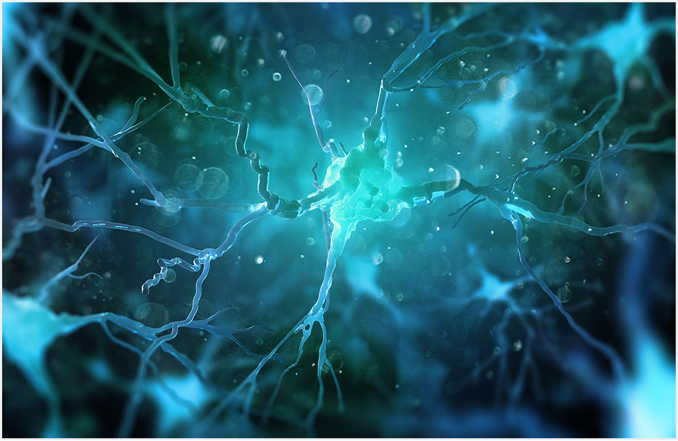
Based on the "two-photon imaging technology" that records detailed events at the micro level of the brain, we have developed various technologies for live observation of the living brain using mice, which are the same mammalian model animals as humans, and have tackled the problem of the "mind". In addition, by developing sensing devices that can be inserted into the brain, we have realized the measurement of new physical indicators and made it possible to observe various phenomena hidden in the brain that no one has been able to investigate yet.
We are now aiming to establish a new platform for in vivo nano quantum imaging in addition to the imaging technology based on the classical quantum "light". Through the development of new technologies to introduce quantum sensors into the above imaging technologies, we will elucidate new "mental" mechanisms, develop observation technologies for brain organoids for application to regenerative medicine, and promote the development of fundamental technologies to support the 100-year age of life. We will continue to develop fundamental technologies to support the age of 100 years of life.
References
Creation of Cell Measurement and Manipulation Technology by Fusion of Nano Quantum Sensor and Microfluidics and Quantum Life Science Application

The measurement technology of "physicochemical quantities" using nano-quantum sensors is making remarkable progress, and is expected to expand to further basic biology research and medical applications. However, the current method involves selecting suitable cells on a culture dish and cutting them off at a certain point in time for measurement, which poses issues of measurement timeliness, throughput, and reproducibility. In addition, the culture dish is far from the actual biological environment, and a space that can provide a space size and chemical environment similar to the biological environment is required to correctly measure cell activity.
Microfluidics technology can control the spatial and chemical environment and align single cells through manipulation by using the micro-space created on the chip. Research is also advancing to create models of organs and tissues on chips by taking advantage of microfluidics' microscopic nature and fluidic networks. By constructing an on-chip cellular measurement space that takes advantage of the microfluidic features described above, we are developing technologies to establish physicochemical measurement techniques with improved timeliness, throughput, and reproducibility for medical applications, and to develop model measurements that mimic the biological environment.